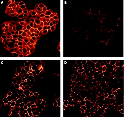
Figure 2 Immunofluorescent images of HT29 cells on confocal microscopy. (A,B) Stained for β‐catenin. (A) Untreated cells, (B) after coculture for 24 h with Helicobacter pylori strain 60190. Decreased membranous staining intensity is seen in (B). (C,D) Stained for E‐cadherin. (C) Untreated cells, (D) after coculture with H pylori strain 60190. No changes can be seen in the staining pattern or intensity.

An official website of the United States government
Here's how you know
Official websites use .gov
A
.gov website belongs to an official
government organization in the United States.
Secure .gov websites use HTTPS
A lock (
) or https:// means you've safely
connected to the .gov website. Share sensitive
information only on official, secure websites.
