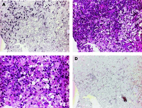
Figure 3 Localisation and morphological identification of B19‐positive cells comparing B19 immunohistochemistry (IHC) and respective haematoxylin and eosin (H&E) staining of Rosai–Dorfman disease (RDD)‐affected nasal mucosa tissue (case 3). (A) B19 VP1/VP2 IHC using alkaline phosphatase‐mediated nitroblue tetrazolium staining (purple black) showing an accumulation of multiple B19‐positive cells with lymphocyte morphology (×20). (B) H&E staining of the same tissue area as shown in (A), localising B19 detection in an area of dense lymphoplasmocytic infiltration and predominantly in a B19‐negative area with neighbouring histiocytic cells (×20). (C) H&E‐stained tissue at higher magnification (×40) showing lymphophagocytosis of histiocytes at the border to the lymphoplasmocytic infiltration. (D) IHC negative control of the respective RDD tissue omitting the B19 VP1/VP2 antibody, showing no specific staining of cells (×20).

An official website of the United States government
Here's how you know
Official websites use .gov
A
.gov website belongs to an official
government organization in the United States.
Secure .gov websites use HTTPS
A lock (
) or https:// means you've safely
connected to the .gov website. Share sensitive
information only on official, secure websites.
