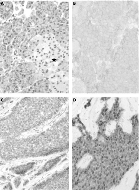
Figure 1 β‐catenin expression in normal and neoplastic gastroenteropancreatic endocrine cells. In normal endocrine pancreatic cells (A), β‐catenin is expressed at low levels all over the islet of Langerhans (*) as compared with adjacent exocrine pancreatic cells. In endocrine tumours, β‐catenin usually retains a membranous distribution; the expression level is either homogeneous (B) or heterogeneous (C) within the tumour. The apparent up regulation of β‐catenin at the periphery of tumour nodules (C) can be seen. Nuclear accumulation (D) of immunoreactive β‐catenin is readily visible in this case. Immunoperoxidase 250×.

An official website of the United States government
Here's how you know
Official websites use .gov
A
.gov website belongs to an official
government organization in the United States.
Secure .gov websites use HTTPS
A lock (
) or https:// means you've safely
connected to the .gov website. Share sensitive
information only on official, secure websites.
