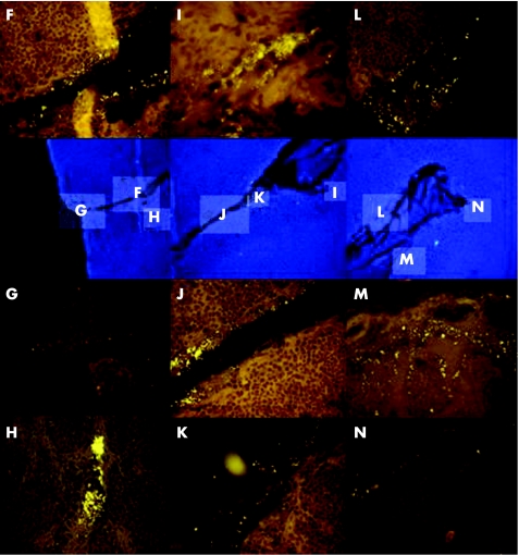
Figure 2 The blue 4′,6‐diamidino‐2‐phenylindole (DAPI)‐stained images in the second row of the figure show at low magnification (×100) a deep fissure crossing the tonsil. The indicated regions are shown at higher magnification relative to DAPI image after hybridisation with the Eub338‐Cy3 (orange) probe. Bacteria fill the fissure (F–N) and infiltrate the adjacent parenchyma in fissure branches of secondary order (H,M,N).

An official website of the United States government
Here's how you know
Official websites use .gov
A
.gov website belongs to an official
government organization in the United States.
Secure .gov websites use HTTPS
A lock (
) or https:// means you've safely
connected to the .gov website. Share sensitive
information only on official, secure websites.
