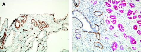
Figure 14 Basal cell markers/α‐methylacyl‐–coenzyme A racemase (AMACR) cocktails with one (A) and two (B) chromogens. (A) p63/AMACR cocktail immunohistochemical stain: p63 is detected in the nuclei of basal cells in benign glands (below), compared with granular cytoplasmic AMACR stain, with apical luminal accentuation (above) in glands of adenocarcinoma, which lack p63. (B) 34βE12/p63/AMACR cocktail: 34βE12 and p63 antibody binding to basal cells is indicated by brown stain, whereas red stain corresponds to AMACR detection. AMACR‐positive, basal‐cell‐negative adenocarcinoma is on the right, whereas benign, AMACR‐negative, basal‐cell‐positive glands are on the left.

An official website of the United States government
Here's how you know
Official websites use .gov
A
.gov website belongs to an official
government organization in the United States.
Secure .gov websites use HTTPS
A lock (
) or https:// means you've safely
connected to the .gov website. Share sensitive
information only on official, secure websites.
