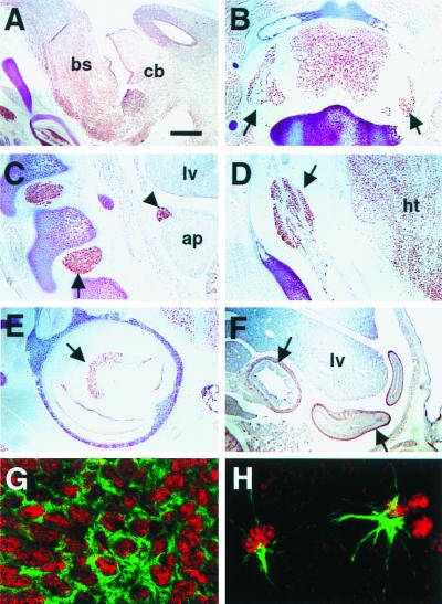
Figure 3.
Distribution of brPTB immunoreactivity in rat and mouse tissues. (A) Sagittal section from E16 rat brain stained with anti-brPTB polyclonal antibody shows strong reactivity with most cells in brain including cerebellum (cb) and brainstem (bs). (B) brPTB immunoreactivity in adult rat spinal cord and dorsal root ganglia (arrows). (C and D) brPTB expression in the developing peripheral nervous system. Immunostaining of E16 rat sagittal sections indicate that brPTB expression is strong in embryonic dorsal root ganglia (C, arrow), trigeminal ganglia (D, arrow), developing adrenal medulla (C, arrowhead) and hypothalamus (D, ht); however, it is very low in liver (C, lv) and adrenal primordium (C, ap). (E) brPTB staining in P5 mouse cochlea. Note the intensive staining in cochlear spiral ganglion cells (arrow). (F) brPTB expression in the ganglion cells of the small intestine (E16 rat embryo). (G) Confocal image of immunofluorescence double exposure of MAP2 (green) and brPTB (red) in a sagittal section of E16 rat cortex. (H) Confocal image of immunofluorescence double exposure of glial fibrillary acidic protein (green) and brPTB (red) in a sagittal section of adult rat cerebellum. Control sections stained with preimmune serum or with brPTB antibody preadsorbed with brPTB fusion protein were negative (data not shown). (Scale bar: 80 μm in A–E; 160 μm in F; 3.4 μm in G; 2.3 μm in H.)

