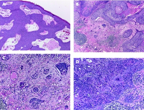
Figure 1 (A) Poroma: multiple epidermal attachments with downgrowth into the dermis. (B) Porocarcinoma, with clear cell change. (C) Higher power of porocarcinoma demonstrating cellular atypia, tumour necrosis, infiltration and budding into adjacent desmoplastic stroma. (D) In situ component adjacent to the invasive porocarcinoma depicted in (B) and (C).

An official website of the United States government
Here's how you know
Official websites use .gov
A
.gov website belongs to an official
government organization in the United States.
Secure .gov websites use HTTPS
A lock (
) or https:// means you've safely
connected to the .gov website. Share sensitive
information only on official, secure websites.
