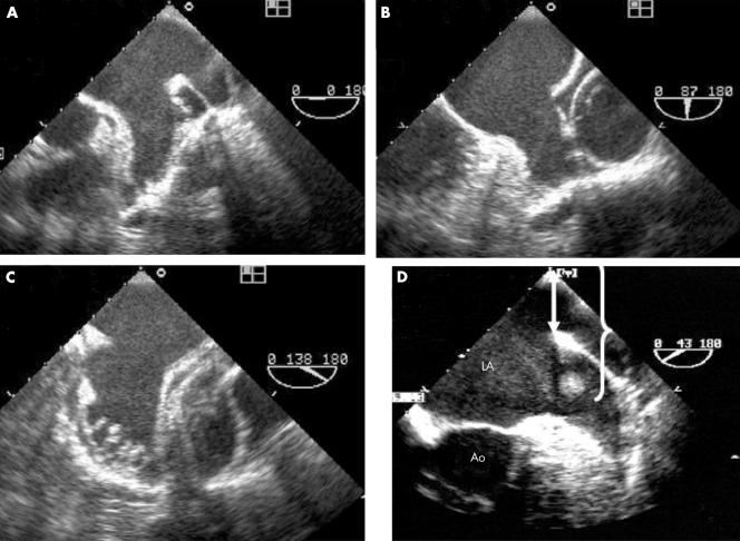
Figure 1 Multiplane transoesophageal echocardiography (TOE) example of (A, B, C) a complete and systematic examination of a left atrial appendage (LAA) without thrombosis and (D) of an LAA artefact. Panels A, B, and C show a multilobed appendage displayed at 0°, 87°, and 138°, respectively. Panel D shows an image inside the LAA that is exactly twice as far from the transducer as from the anatomical interface produced by the bend between the LAA and left pulmonary vein (arrow and brackets). Ao, aorta; LA, left atrium.

An official website of the United States government
Here's how you know
Official websites use .gov
A
.gov website belongs to an official
government organization in the United States.
Secure .gov websites use HTTPS
A lock (
) or https:// means you've safely
connected to the .gov website. Share sensitive
information only on official, secure websites.
