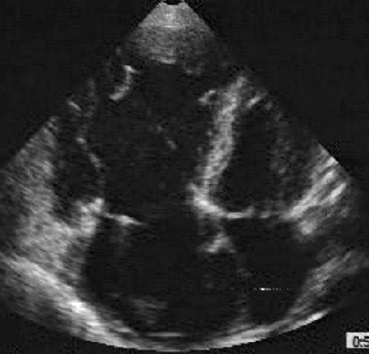
Figure 10 Apical four chamber view from a patient with a severe form of ARVC/D. In contrast with the patient in fig 9 where the aneurysm was very localised, here the entire RV is dilated and poorly contracting.

Figure 10 Apical four chamber view from a patient with a severe form of ARVC/D. In contrast with the patient in fig 9 where the aneurysm was very localised, here the entire RV is dilated and poorly contracting.