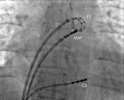
Figure 3 Electrical isolation of the left superior pulmonary vein. A fluoroscopic image of the heart viewed in the anterior posterior projection. The pulmonary vein catheter (PV) has 14 electrodes in a spiral and is positioned in the left superior pulmonary vein recording the electrograms inside the vein. The ablation catheter (MAP) is at the ostium of the vein where it joins the left atrium. Through this catheter radiofrequency energy is delivered to ablate the connections between the left atrium and pulmonary vein until electrical isolation of a pulmonary vein is achieved. A catheter is also seen in the coronary sinus (CS) which can be used to pace to separate the pulmonary vein potential form the far field left atrial potential.

An official website of the United States government
Here's how you know
Official websites use .gov
A
.gov website belongs to an official
government organization in the United States.
Secure .gov websites use HTTPS
A lock (
) or https:// means you've safely
connected to the .gov website. Share sensitive
information only on official, secure websites.
