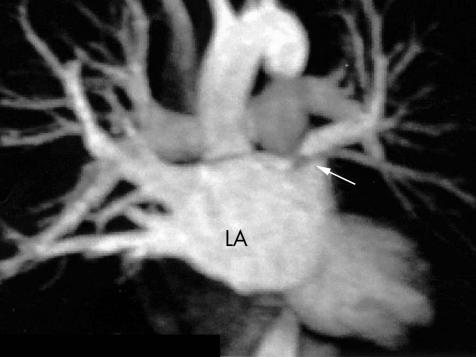
Figure 6 A contrast enhanced magnetic resonance image of the left atrium (LA) showing a severe ostial stenosis of the left superior pulmonary vein. The body of the left atrium is viewed in the anteroposterior (AP) projection. The right superior and inferior pulmonary veins are visible and the arrow indicates the stenosis. This patient was asymptomatic; however, reduced perfusion to the left lung was demonstrated by a VQ scan and a successful balloon angioplasty of this vessel was performed.

An official website of the United States government
Here's how you know
Official websites use .gov
A
.gov website belongs to an official
government organization in the United States.
Secure .gov websites use HTTPS
A lock (
) or https:// means you've safely
connected to the .gov website. Share sensitive
information only on official, secure websites.
