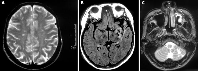
Figure 1 Magnetic resonance images of the brain. (A) T2 weighted image of a 35 year old female patient with Eisenmenger's syndrome, who had no history of stroke but had chronic headache as well as exercise intolerance. Note the multiple small bilateral lesions with high intensity in the centrum semiovale. The packed cell volume was 63%. (B) Fluid attenuated inversion recovery image of a 25 year old female patient with corrected transposition of the great arteries, ventricular septal defect, and hypoplastic pulmonary arteries, who had no history of stroke or any hyperviscosity related symptoms. Note the old infarction and atrophic changes in the left temporal lobe. The packed cell volume was 61%. (C) T2 weighted image of a 23 year old male patient with tetralogy of Fallot and extremely hypoplastic pulmonary arteries, who had chronic headache, dizziness, and taste disorder. Note the old infarction in the left cerebellar hemisphere. The packed cell volume was 74%.

An official website of the United States government
Here's how you know
Official websites use .gov
A
.gov website belongs to an official
government organization in the United States.
Secure .gov websites use HTTPS
A lock (
) or https:// means you've safely
connected to the .gov website. Share sensitive
information only on official, secure websites.
