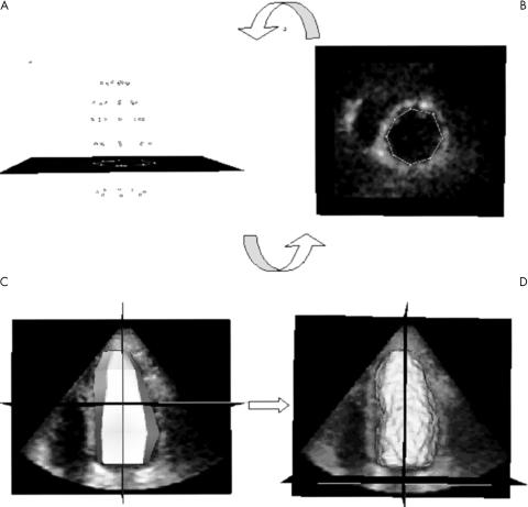
Figure 2 (A, B) Point verification and correction and (C, D) endocardial surface detection. The initialised points (A) are shown in each short axis plane (B) for verification and correction if required. The endocardial surface (C) obtained by joining these points is the initial condition for the three dimensional endocardial detection algorithm, which results in the calculated LV endocardial surface (D).

An official website of the United States government
Here's how you know
Official websites use .gov
A
.gov website belongs to an official
government organization in the United States.
Secure .gov websites use HTTPS
A lock (
) or https:// means you've safely
connected to the .gov website. Share sensitive
information only on official, secure websites.
