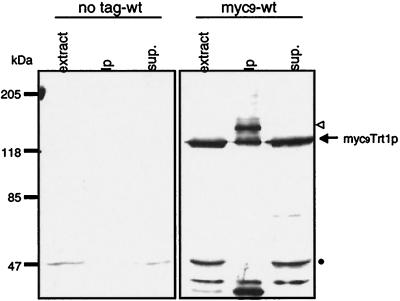
Figure 3.
Detection of Cmyc9Trt1p fusion protein in cell extracts. Immunoblot of extracts prepared from a wild-type S. pombe strain with untagged Trt1p (CF199) and a strain expressing C-terminal myc9-tagged Trt1p (CF830). Crude extract (50 μg of total protein), immunoprecipitation with monoclonal anti-c-myc-antibody bound to agarose beads, and the supernatant thereof were separated by SDS/PAGE. Tagged proteins were detected by immunoblotting using a rabbit polyclonal anti-c-myc primary antibody (Santa Cruz Biotechnology) and peroxidase-conjugated goat anti-rabbit IgG secondary antibody (Boehringer Mannheim). Putative bands for the myc9Trt1p fusion protein are indicated by an arrow and open triangle (see text). The band at ≈50 kDa indicated by a dot is unrelated to Trt1p.

