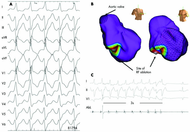
Figure 8 Non‐contact mapping of an idiopathic left ventricular tachycardia with right bundle branch block, leftward axis (A). The multiple electrode array (MEA) was placed in the left ventricle. The MEA is part of the non‐contact mapping system (EnSite 3000; Endocardial Solutions, Inc). The system permits mapping of a single complex. The MEA, which is filled with a contrast–saline medium, is positioned in the left ventricle (right/left anterior oblique views). The system calculates electrograms from 3000 endocardial points simultaneously by reconstructing far‐field signals. Non‐depolarised myocardium is shown in purple in this three dimensional isopotential map (B). The map also shows the site of earliest depolarisation (white circle). At the distal part of the left posterior fascicle, radiofrequency ablation almost immediately terminated the VT (C), which, thereafter, was no longer inducible.

An official website of the United States government
Here's how you know
Official websites use .gov
A
.gov website belongs to an official
government organization in the United States.
Secure .gov websites use HTTPS
A lock (
) or https:// means you've safely
connected to the .gov website. Share sensitive
information only on official, secure websites.
