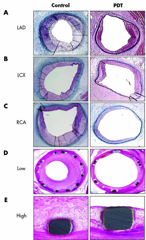
Figure 2 Representative histological appearances of balloon overstretch‐injured (BI) and stented arteries subjected to photodynamic therapy (PDT) and control. Porcine coronary arteries were subjected to BI followed by PDT. (A) Left anterior descending artery (LAD); (B) left circumflex artery (LCX); (C) right coronary artery (RCA). Animals were killed 14 days later. Porcine coronary arteries were subjected to stenting followed by PDT. Animals were killed 30 days later. (D) Low magnification 20×; (E) high magnification 200×. Sections of perfusion‐fixed arteries were stained with haematoxylin and eosin.

An official website of the United States government
Here's how you know
Official websites use .gov
A
.gov website belongs to an official
government organization in the United States.
Secure .gov websites use HTTPS
A lock (
) or https:// means you've safely
connected to the .gov website. Share sensitive
information only on official, secure websites.
