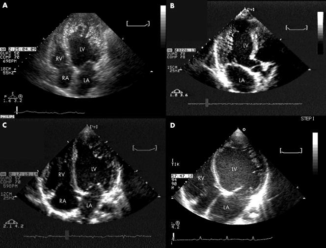
Figure 1 Representative examples at two‐dimensional echocardiography of the spectrum of left ventricular (LV) functional and morphological impairment in isolated ventricular non‐compaction. (A) Non‐compaction involving the entire apex in a patient with normal end diastolic LV volume (with an overall picture that can mimic apical hypertrophic or obliterative cardiomyopathy); (B) non‐compaction limited to the posterior wall in a patient with nearly normal end diastolic LV; (C) non‐compaction involving the apicolateral wall in a patient with mildly dilated cardiomyopathy; (D) non‐compaction involving the apicolateral wall in a patient with severely dilated cardiomyopathy. LA, left atrium; LV, left ventricle; RA, right atrium; RV, right ventricle.

An official website of the United States government
Here's how you know
Official websites use .gov
A
.gov website belongs to an official
government organization in the United States.
Secure .gov websites use HTTPS
A lock (
) or https:// means you've safely
connected to the .gov website. Share sensitive
information only on official, secure websites.
