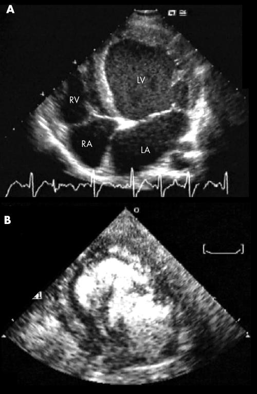
Figure 3 Example of the often decisive role of contrast echocardiography in the diagnosis of isolated ventricular non‐compaction: at two‐dimensional echocardiography in the four‐chamber view, a morphologically ambiguous picture (A) is clarified by use of the echo‐contrast medium (B), which accentuates the true trabecular pattern. LA, left atrium; LV, left ventricle; RA, right atrium; RV, right ventricle.

An official website of the United States government
Here's how you know
Official websites use .gov
A
.gov website belongs to an official
government organization in the United States.
Secure .gov websites use HTTPS
A lock (
) or https:// means you've safely
connected to the .gov website. Share sensitive
information only on official, secure websites.
