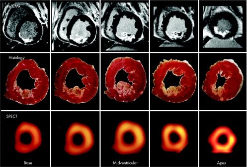
Figure 2 Contrast enhanced cardiovascular magnetic resonance (CeCMR), histology and single photon emission computed tomography (SPECT) images obtained in an animal with a medium sized infarct. There is a nearly perfect match between necrosis defined by histology and ceCMR. Whereas ceCMR allows the exact assessment of the transmural extent of infarction, SPECT defines segments as either viable or non‐viable. Reproduced with permission from Wagner et al.9

An official website of the United States government
Here's how you know
Official websites use .gov
A
.gov website belongs to an official
government organization in the United States.
Secure .gov websites use HTTPS
A lock (
) or https:// means you've safely
connected to the .gov website. Share sensitive
information only on official, secure websites.
