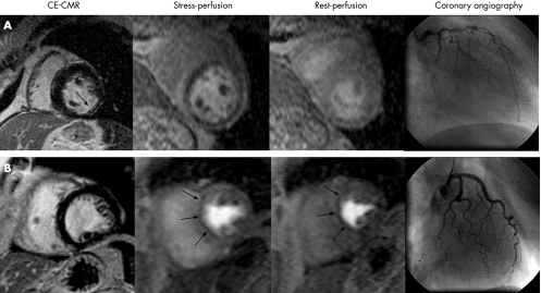
Figure 6 Comprehensive CMR stress perfusion study. The upper panel depicts a positive ceCMR study on the left demonstrating subendocardial infarction in the inferolateral wall (single arrow). Consequently, this patient was ruled positive for CAD (see fig 5, step 1). Adenosine perfusion CMR revealed prominent reversible defects in all coronary artery territories (dark areas in stress perfusion), indicating myocardial ischaemia. Since the perfusion defects were significantly larger than the infarct, viable myocardium is at risk. Coronary angiography demonstrated multivessel disease in this patient, who then underwent successful surgical revascularisation. Panel B displays a patient with a matched stress–rest perfusion defect (arrows) but without evidence of prior MI on ceCMR (left bottom panel). Thus, the perfusion defects must be classified as artefactual and the patient as negative for CAD (see fig 5, step 4). Coronary angiography demonstrated normal coronary arteries.

An official website of the United States government
Here's how you know
Official websites use .gov
A
.gov website belongs to an official
government organization in the United States.
Secure .gov websites use HTTPS
A lock (
) or https:// means you've safely
connected to the .gov website. Share sensitive
information only on official, secure websites.
