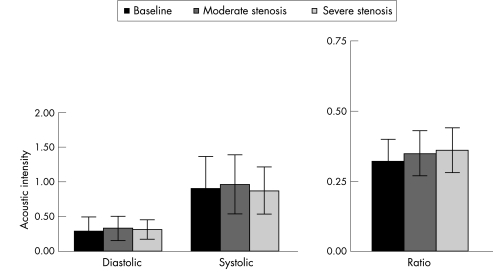
Figure 5 Diastolic and systolic acoustic intensity values and the systolic to diastolic arteriolar blood volume ratio from the collateralised region of the left anterior descending coronary artery bed at baseline and at 2 levels of stenosis in the 12 dogs. There are no differences between these values.

An official website of the United States government
Here's how you know
Official websites use .gov
A
.gov website belongs to an official
government organization in the United States.
Secure .gov websites use HTTPS
A lock (
) or https:// means you've safely
connected to the .gov website. Share sensitive
information only on official, secure websites.
