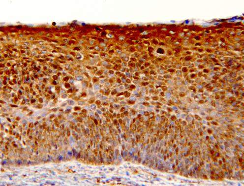
Figure 3 A low‐grade cervical intraepithelial neoplasia 1 lesion, showing intense staining for nm23‐H1 throughout the full thickness of the lesion, being mainly cytoplasmic; many positive nuclei are also present. The underlying stroma is nm23‐H1 negative (immunohistochemistry for nm23‐H1, original magnification ×200).

An official website of the United States government
Here's how you know
Official websites use .gov
A
.gov website belongs to an official
government organization in the United States.
Secure .gov websites use HTTPS
A lock (
) or https:// means you've safely
connected to the .gov website. Share sensitive
information only on official, secure websites.
