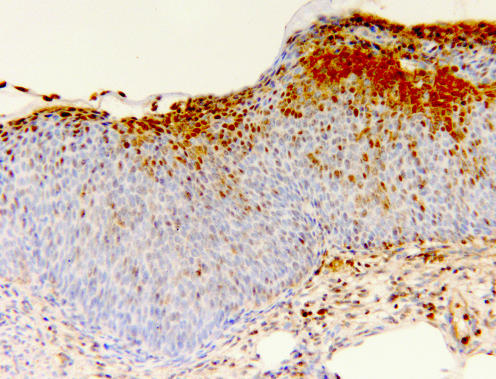
Figure 4 A high‐grade cervical intraepithelial neoplasia 3 lesion, with markedly reduced nm23‐H1 expression. Positive staining is confined to the uppermost layers of the epithelium, whereas the lower two thirds remains entirely nm23‐H1 negative, except for some single scattered positive cells (immunohistochemistry for nm23‐H1, original magnification ×100).

An official website of the United States government
Here's how you know
Official websites use .gov
A
.gov website belongs to an official
government organization in the United States.
Secure .gov websites use HTTPS
A lock (
) or https:// means you've safely
connected to the .gov website. Share sensitive
information only on official, secure websites.
