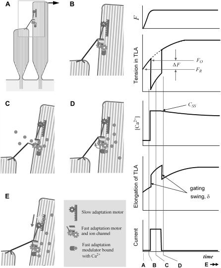
FIGURE 2.
Mechanism of mechanoelectric transduction in the presented model. Our computer model considers the transduction channels to be serially connected to the tip links. The transduction channel and adaptation motor are located at the upper end of the tip link. Although the slow adaptation is not modeled or simulated in this study, it was included in this illustration. (Left) At rest, the channel is closed (A), and the TLA has a resting tension of FR. As the bundle deforms, the tension in the TLA increases (B). When the tension exceeds the opening tension level, FO, the channel opens (C). Due to the gating swing (δ) the tension in TLA drops. The cations flow in due to the resting potential between extracellular and intracellular fluid. As Ca2+ at the site of the fast adaptation modulator increases, the channel closes and the tip link pulls back the taller stereocilium (D). By sliding down the upper end of the tip link (slow adaptation), the tension of TLA is decreased to its resting level (E). (Right) The time response of a single channel from our proposed model. From top to bottom, the applied force to the bundle, tension in the TLA, [Ca2+] at the fast adaptation site, elongation of the TLA, and transduction current of the single channel. The vertical lines A–D correspond to the images labeled A–D to the left. This study simulates for 2 ms after the onset of excitation, before the slow adaptation (E) becomes apparent.

