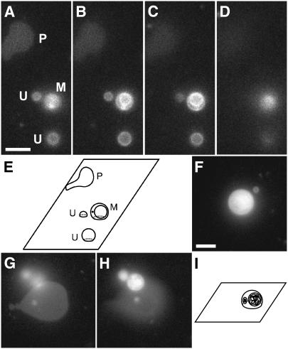
FIGURE 1.
Distinguishing characteristics of planar bilayers, unilamellar vesicles, and multilamellar vesicles. DOPC/DOPG (9:1) GUVs labeled with 0.5 mol % TR-DHPE were adsorbed to a glass coverslip and visualized using Texas Red fluorescence emission. Unless otherwise noted, the focal plane was the glass surface. Scale bar in A and F, 5 μm. (A–D) A series of focal planes of the same coverslip-adsorbed GUV sample, with the focal plane 0, 1, 2, and 4 μm, respectively, above the glass surface. A planar bilayer, two unilamellar vesicles, and a multilamellar vesicle in A are marked P, U, and M, respectively. (E) Schematic illustration of the planar bilayer and the vesicles observed in A–D. (F–H) Rupture of a multilamellar GUV. (F) An adsorbed GUV. (G) The lipid structures in F after GUV rupture. A planar bilayer and the fuzzy outline of additional lipid structures above the surface are visible. (H) The lipid in F after GUV rupture at a focal plane above the surface. The internal lipid structures were identified as vesicles, indicating that the original GUV was multilamellar. The focal plane is 2.7 μm above the glass surface. (I) Schematic illustration of the adsorbed GUV in F. Cut-away views show the multilamellarity.

