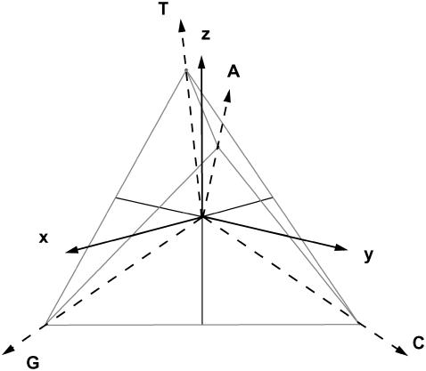
FIGURE 2.
Orthonormal x-y-z base set and tetrahedral DNA-nucleotide set representation. Each of the three axes distinguishes a specific molecular class feature. Purines are distinguished from pyrimidines through x-coordinate. Amino are distinguished from keto through y-coordinate. Weak Watson-Crick hydrogen-bridge binding is distinguished from stronger binding through z-coordinate.

