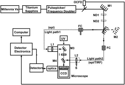
FIGURE 1.
Experimental setup to study picosecond FLIM and FRET using point and imaging detectors in the epifluorescence and TIRF modes. OCFD, optical constant fraction discriminator triggered by laser pulse; M, mirrors; ND, neutral density filters; UV, mercury lamp for steady-state imaging; FC, optic fiber coupler; epi, epifluorescence mode; TIRF, total internal reflection fluorescence mode; L, planar convex lens; I, iris to control the area of excitation of the sample; S, micrometer screw, and CCD, charge-coupled device for steady-state imaging.

