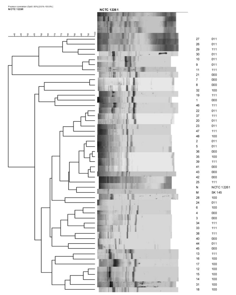
FIGURE 2.


Western immunoblots were run on cell wall extracts of the same isolates tested by ELISA. The blots were developed using antisera against S. mitis biovar 1 strains NCTC 12261T (Figure 2A) and SK 145 (Figure 2B). Antisera to S. oralis strains ATCC 35037T and SK100 were non-reactive in Western blotting. Digital images of the blot strips were compared by cluster analysis using UPGMA. To the right of the strips is shown the identity of the isolate and its phenotype.
