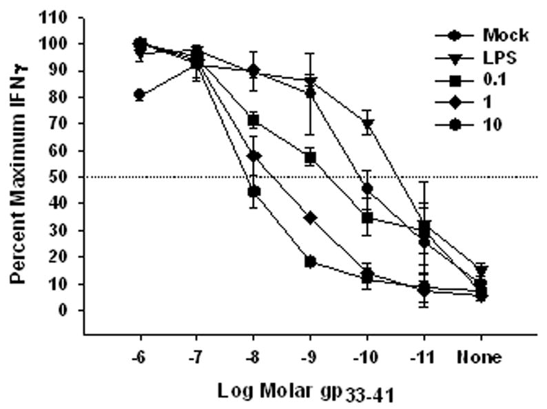
Figure 5. Decreased antigen presentation by VV-infected BMDC.

Mouse BMDC were mock-treated, treated with LPS, or infected with VV (MOI = 0.1, 1, or 10) for 18 hours and then pulsed with varying concentrations of gp33-41. Following washing to remove unbound peptide, BMDC were co-cultured with a gp33-41-specific CTL line for 5 hours in the presence of GolgiPlug. IFNγ production was determined by FACS analysis and expressed as percent maximal IFNγ production. Data are the average of 3 independent experiments.
