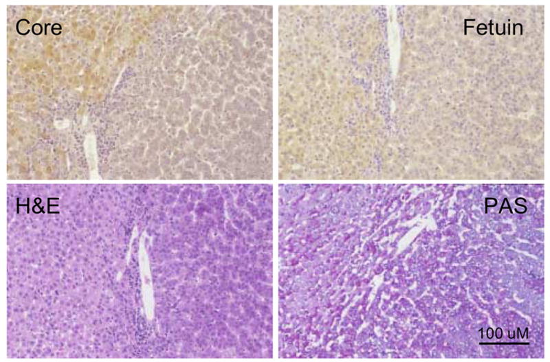
Figure 2. Lack of WHV core antigen expression in HCC.

Liver tissue sections showing areas of WHV core antigen positive hepatocytes adjacent to areas of HCC in woodchuck 3202. Adjacent liver sections were stained for detection of WHV core antigen and fetuin B protein or stained by H&E and PAS as described in Materials and Methods. In each panel hepatocytes with normal morphology and detectable WHV core antigen expression were located on the left hand side of the field. The area showing HCC contains characteristic clusters of hepatocytes, 4 cells thick, and disruption of the hepatic plates. Magnification 160x. Bar = 100 uM.
