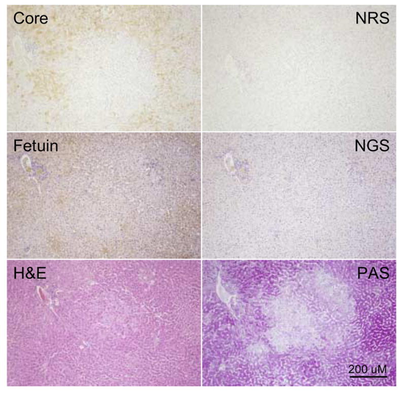
Figure 4. Lack of WHV core antigen expression in amphophilic FAH.

Liver sections showing an amphophilic WHV core antigen negative FAH from woodchuck 307 (26 months of age). Adjacent sections were immuno-stained for detection of WHV core antigen using NRS as a control, or fetuin B protein, using NGS as a control, or stained by H&E or PAS as described in Materials and Methods. The prominent portal tract in the upper left hand corner of each field was used to locate the FAH. The amphophilic FAH, which has some basophilic cells present giving it a mixed cell phenotype, has undetectable levels of WHV core antigen, and reduced but variable levels of fetuin B protein. The FAH has some morphological changes seen on H&E and has lower levels of glycogen leading to lighter staining by PAS. Magnification 80x. Bar = 200 uM.
