
Figure 6. Foci of WHV core antigen negative hepatocytes not morphologically distinguishable as FAH.
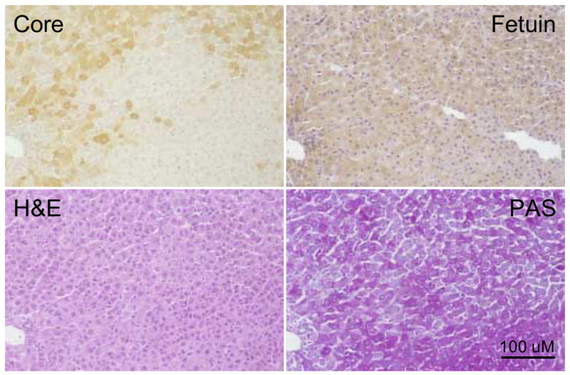
Adjacent liver sections showing a morphologically normal focus of WHV core antigen negative hepatocytes (woodchuck 4961, 19 months of age) subjected to fetuin B protein staining, H&E or PAS. The portal tract in the lower left hand corner has been used to locate the same area in each adjacent liver section. In this case the focus of WHV core antigen negative hepatocytes is not morphologically different from surrounding hepatocytes as judged by fetuin B protein detection or H&E or PAS staining. Magnification 160x. Bar = 100 uM.