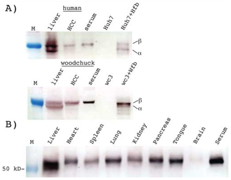
Figure 9. Evidence for liver specific expression of fetuin B protein by western blotting.

A) Fetuin B expression was determined using human and woodchuck liver, HCC and serum samples, and Huh7 and wc3 hepatoma cell lines transfected with pHuFetuin-B (Huh7+Hfb) and pWcFetuin-B (wc3+Wfb) respectively. Liver and HCC samples were homogenized, then protein concentrations were determined and the equivalent of 50 μg of human and 100 μg of woodchuck tissue was loaded per lane for SDS polyacrylamide gel electrophoresis. 0.2 ul of human and woodchuck serum and 10 μg of Huh7 or wc3 cell lysate was also loaded. Western blots were probed with anti-human fetuin B antibodies, as described in Materials and Methods. Both forms of fetuin B protein (α and β) were detected in normal human and woodchuck liver tissue and transfected Huh7 and wc3 cells. Only the β form was detected in HCC and serum. B) Western blot analysis confirming the presence of the β form of fetuin B protein in each woodchuck tissue analyzed. The α form of fetuin B was only detected in the liver.
