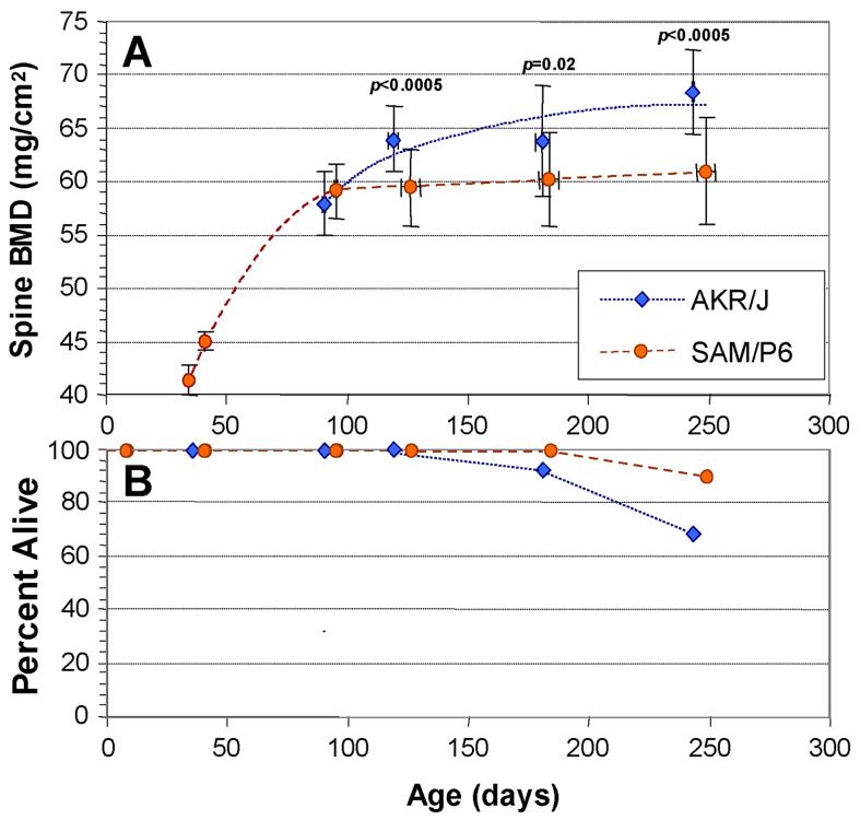
Figure 1.
Age-dependence of spinal BMD in the AKR/J and SAMP6 mouse strains. Bone mineral density (BMD) was determined for the spines of individual, transponder identified mice of each strain at the ages indicated (A). Mice were excluded if there was superposition of transponders with the spinal column; the remaining assay cohorts comprised 24 AKR mice and 23 SAMP6 mice. Error bars show standard deviations (SDs) for both age (x-axis) and spine BMD (y-axis). Significances are given for 2-tailed t-tests (Fisher-Behrens test for samples of unknown or unequal variance), comparing spine BMDs of strains at 4, 6 and 8 months. Survivals of the two cohorts are shown in the lower panel (B); at 8 months of age, 16 AKR and 21 SAMP6 mice remained.

