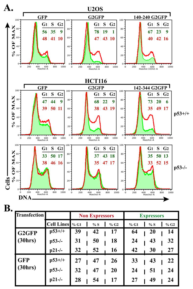
Fig. 11. Ectopic cyclin G2 expression induces a p53-dependent cell cycle arrest that does not require p21 expression.

(A) The DNA content (Hoechst 33342 labeled) of intact, (propidium iodide negative) live cells transfected with expression plasmids for full length, truncated cyclin G2-GFP, or control GFP was analyzed by flow cytometry. Amounts of DNA (Hoechst) of the GFP expressing cells are shown as histograms (shaded in green) overlaid onto the DNA histogram of the nonexpressing population (red line) from the same transfection reaction. Nonexpressing and GFP expressing cells in each transfected population were collected and analyzed simultaneously by the cytometer. The percentage of cells in the G1, S and G2+M phases, as determined using the FlowJo Watson Pragmatic cell cycle analysis program, is shown at the right in color-coded font corresponding to the indicated histogram. [Similar results were obtained applying the Dean-Jett-Fox cell cycle modeling algorithm and similar results were obtained in three independent experiments for each cell line.] Top panels show analysis of U2OS cells transfected with GFP (left) full length cyclin G2-GFP (middle) and 140–240 cyclin G2-GFP (right). Middle and bottom panels illustrate the analysis of p53+/+ and p53−/− HCT116 cells, respectively, expressing GFP (left), cyclin G2-GFP (middle) and 142–344 cyclin G2-GFP (right). (B) Table shows the percentage of cells in each cell cycle phase, comparing GFP and G2-GFP expressors to nonexpressors, in p53+/+, p53−/− and p21−/− isogenic HCT116 cells as determined by the FlowJo Watson Pragmatic cell cycle analysis program. Note the intermediary effect on cell cycle arrest with ectopic expression of cyclin G2 in p21 null cells.
