
Fig. 2. Endogenous and ectopic cyclin G2 distributes to detergent-resistant insoluble protein fractions.
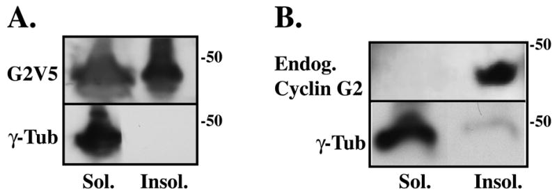
(A, B) Immunoblot analysis of soluble (Sol.) and insoluble (Insol.) proteins fractionated from cyclin G2V5His transfected (A) and nontransfected (B) U2OS cells. Cells were harvested, washed in PBS, and lysed in PHEM buffer containing 1% Triton X-100. A low speed 300 g centrifugation step separated soluble proteins from the insoluble protein pellet. Soluble proteins present in the supernatant were precipitated with methanol and pelleted. The soluble and insoluble protein pellets were washed in PHEM buffer, solubilized in sample buffer and an equivalent volume of each fraction was loaded onto a SDS-PAGE gel. Immunoblot analysis using rabbit anti-cyclin G2 and mouse anti-γ-tubulin shows the distribution of ectopic G2V5 (A) and endogenous G2 (B) primarily in the detergent resistant insoluble fraction relative to γ-tubulin, which distributes to the soluble fraction.