
Fig. 7. Centrosomal localization of GFP-tagged AKAP450 C-terminus dissociates endogenous cyclin G2 from the centrosome.
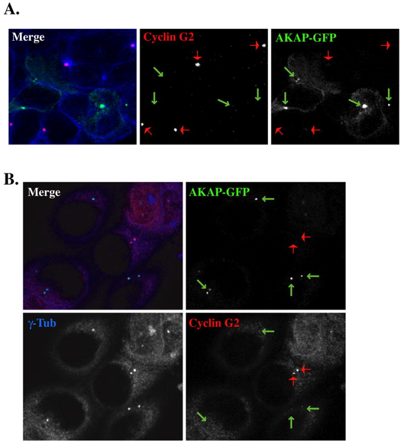
Micrographs of 0.3 μM optical sections from AKAP-GFP transfected U2OS (A) or MCF-7 (B) cells, fixed with MeOH, and immunostained with antibodies against cyclin G2 (red) in (A and B), and α- (in A) or (in B) γ-tublin (blue). Shown in merge fields are the pseudo-colored fluorescence signals of the single channels black and white. The arrows indicate the centrosomal localization of AKAP-GFP (green) and cyclin G2 (red). Note the absence of centrosomal cyclin G2 in cells expressing AKAP-GFP at MTOCs/ centrosomes.