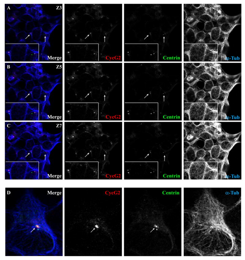
Fig. 8. Endogenous cyclin G2 localizes with only one centriole of the interphase centrosome, chiefly the MT-associated (mature) centriole.

Shown are confocal immunofluorescence micrographs of U2OS cells transfected with GFP-tagged centrin and stained for endogenous cyclin G2 and α-tubulin. (A-C) Serial 0.3 μM sections (Z3, 5, and 7) of the same field show the localization of endogenous cyclin G2 staining relative to two GFP-centrin-labeled centriole pairs (arrows). The inset at the lower left shows a higher magnification of this area. Note that cyclin G2 colocalized with only one of the two GFP-centrin-positive centrioles in a pair. The cyclin G2-reactive centrioles were typically close to the center of radial MT arrays as is typical for MCs, whereas DCs are usually off this center. (D) Zoomed-in image of a GFP-centrin positive MTOC from a cell in another field showing colocalization of cyclin G2 with only one of two centrioles. Note that the larger of two GFP-labeled centrioles is at the center of a MT array and co-stained by anti-cyclin G2 antibodies, but the smaller centriole off to the outside edge of the MTOC does not co-stain with cyclin G2 antibodies. Similar results were observed in several other independent experiments.
