Figure 2.
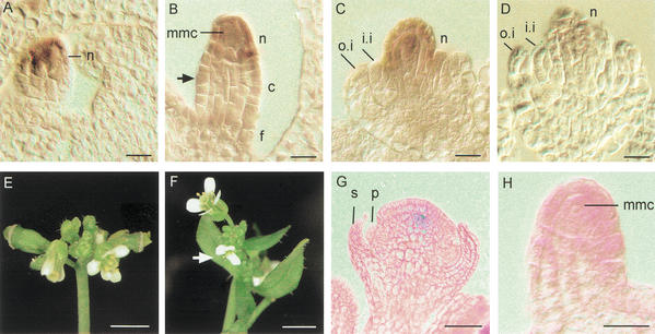
WUS mRNA expression in ovules and characterization of CLV1::WUS plants. (A–D) In situ hybridization to tissue sections of various stages of wild type ovules. Signal is detected as brown color. (A) WUS mRNA is detected in young ovules in the nucellus. (B) At stage 2-II, when the inner integuments are initiated (arrow) a strong signal is detected in the nucellus of the ovule while no mRNA expression is observed in the chalaza and the funiculus. (C,D) WUS mRNA expression becomes weaker during the outgrowth of integuments at stages 2-IV (C) to 2-V (D). (E,F) Phenotype of CLV1::WUS (E) and CLV1::WUS; wus (F) inflorescences. In F, the arrow points at a flower resembling those found in wus mutants, whereas the top flower contains a gynoecium. (G,H) Expression of the linked CLV1::GUS reporter gene in a floral meristem (G) and a young ovule primordium of CLV1::WUS; wus plants. GUS activity is detected as blue colour in a central cell population of the floral meristem (G). No signal is obtained in the ovule primordium (H). (c) Chalaza; (f) funiculus; (i.i.) inner integument; (mmc) megaspore mother cell; (n) nucellus; (o.i.) outer integument; (p) petal primordium; (s) sepal primordium. Bars, 10 μm (A–D,H), 3 mm (E,F), and 30 μm (G).

