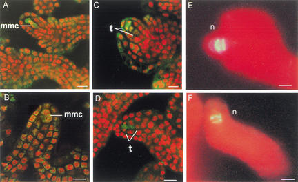
Figure 4.
Meiosis in CLV1::WUS and wus ovules. (A–D) Confocal microscopy of CLV1::WUS and wus ovules. Nuclei are detected in red, cytoplasm appears green. (A,B) In CLV1::WUS (A) and wus (B) ovules two nuclei are visible in the megaspore mother cell. (C,D) Tetrad formation is evident in CLV1::WUS (C) and wus (D) ovules. (E,F) Dark-field microscopy of CLV1::WUS and wus ovules. Ovules were stained with aniline blue to detect callose accumulation, which is indicative of meiotically dividing cells. Autofluorescence of the tissue is detected in red. Callose accumulation is shown as bright color. In both CLV1::WUS (E) and wus (F) ovules, two bands of callose accumulation are detected. (mmc) Megaspore mother cell; (n) nucellus; (t) tetrad. Bars, 10 μm.

