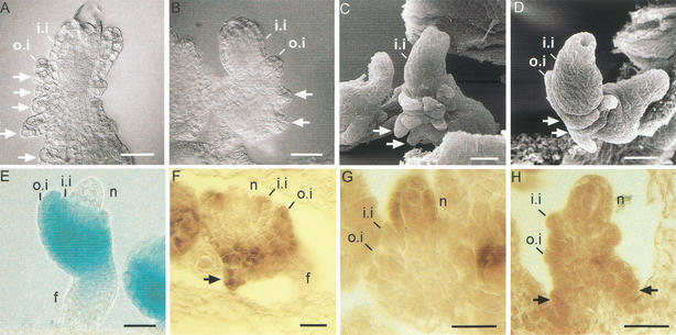
Figure 6.
Ectopic expression of WUS. (A–D) Ovules of dexamethasone induced ANT::WUS-GR plants. DIC microscopy images (A,B) and SEM images (C,D) showing the nucellus enclosed by the inner integument. The outer integument does not grow beyond initial stages. Integument-like structures (arrows) are visible below the normal inner and outer integuments. (E) Expression of an ANT::GUS reportergene in wild-type plants. GUS activity is shown as blue color marking the chalaza including the inner and outer integument. (F) ANT mRNA expression in ovules of induced ANT::WUS-GR plants. ANT mRNA is detected in the chalaza and additionally in the newly formed ectopic structure (arrow). (G,H) WUS mRNA expression. (G) Control plant. WUS mRNA signal is restricted to the nucellus. (H) Ovule of induced ANT::WUS-GR plant. WUS mRNA expression is visible throughout the ovule primordium, including the region that forms ectopic organs (arrows). (f) Funiculus; (i.i.) inner integument; (n) nucellus; (o.i.) outer integument. Bars 20 μm.

