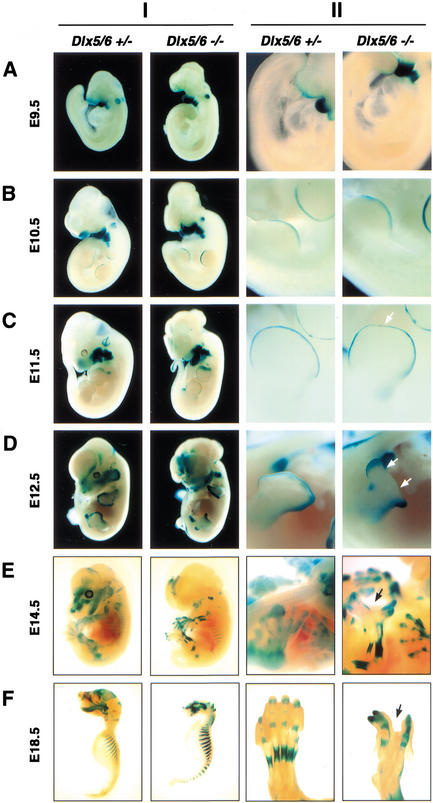
Figure 2.
Embryonic Dlx5 and Dlx6 expression revealed by whole-mount β-galactosidase staining of heterozygous and homozygous Dlx5/6 mutant embryos (Column I), with higher magnifications of limbs from the same embryos (Column II). (A) Dlx5/6 is strongly expressed in the first and second branchial arches and the otic pit, with less prominent expression in the tail and forelimb buds at E9.5. (B) Expression becomes apparent in the AER of both fore- and hindlimb buds by E10.5. (C) At E11.5, reduction in expression in the frontonasal prominence and medial AER (arrow) initiates. (D) Expression in the medial AER deteriorates by E12.5 in Dlx5/6−/− embryos. Arrows in the right-hand column indicate the loss of medial tissue and digits in the Dlx5/6−/− embryos. (E) Expression within all developing bones becomes apparent by E14.5, at which time the SHFM phenotype becomes recognizable in null mutants. (F) At E18.5, craniofacial and skeletal expression remains strong. Calvarial and mandibular expression is noticeably lost in Dlx5/6−/− embryos owing to the absence of these tissues.

