Figure 4.
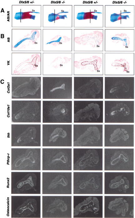
Histological and molecular examination of the ossification of the scapula of heterozygous and Dlx5/6 null embryos at E16.5. (A) AB/AR staining of whole-mount scapulas shows the absence of ossification of the spine of scapula (Ss) and a minimal ossification within the center of the scapula, compared to heterozygous embryos. Vertical lines indicate the approximate area from which comparable serial sections were taken for each column of results. (B) In more proximal sections, AB-stained Dlx5/6−/− scapulas appear to lack both hypertrophic chondrocytes and a von Kossa (VK)-positive calcium matrix. (C) In contrast to control embryos, Col2a1 expression remains strong and Col10a1 appears to be increasing in the Dlx5/6 null sections. In addition, Ihh downregulation appears to be delayed in Dlx5/6 null sections, whereas Pthrp-r expression is unaffected. Runx2 expression appears normal in the perichondrium, but reduced in the chondrium of the Dlx5/6 null sections. Osteocalcin expression is completely absent in the Dlx5/6 null sections. In more distal sections, calcified hypertrophic chondrocytes are now apparent. Col2a1 expression is now almost absent in the Dlx5/6 null sections, and Col10a1 expression is strong. At this level, Runx2 appears normal in the Dlx5/6 null sections and Osteocalcin expression remains absent.

