Figure 6.
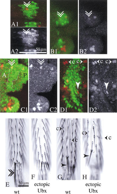
In a regime of prolonged ectopic Ubx expression, neural markers disappear from the apical bristle precursors on T2. The adult flies show selective loss of the apical bristle. (A–D) Confocal images of the apical bristle precursors in prepupal legs with ectopic Ubx expression (Ubx isoform Ia) driven by dpp-Gal4 in the neu-lacZ background. Precursors are marked by anti-β-galactosidase staining (red), and Ubx expression is shown (green). Double arrowhead marks the apical bristle precursors, arrowhead marks the preapical bristle precursors. (A) Second leg about 3 h APF. Second-order precursors for the apical bristle are present within the domain of ectopic Ubx expression. (A2) shows the Ubx channel. (B) Second leg 5–6 h APF. Very low levels of anti-β-galactosidase staining remain in the apical bristle precursors (see red channel in B2). Anti-β-galactosidase staining is also reduced in the apical bristle precursors of the third leg of this individual (C). The stripe of ectopic Ubx expression in the third leg (white outlines in C1) compares to Ubx levels in the posterior third leg (marked P), exceeding levels in the anterior third leg (marked A). (D) In the dorsal second leg, the stripe of ectopic Ubx expression occupies most of the width between the chemosensory precursors (small arrowheads accompanied by the letter c). (E) Ventral aspect of the distal tibia of an adult wild-type fly and (F) of a fly from the above regime of ectopic Ubx expression—the apical bristle is missing from the ventral side of the tibia. From the dorsal domain of ectopic Ubx expression (D) the preapical bristle (arrowhead in H) and mechanosensory microchaetes on the dorsal leg (circle in H) arise normally. (G) Dorsal aspect of the distal tibia of a wild-type fly.

