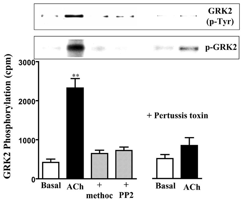
Figure 5. Phosphorylation of GRK2 by c-Src.

Gastric smooth muscle cells labeled with 32P were incubated with ACh (0.1μM) in the presence of absence of methoctramine (0.1 μM) or PP2 (1 μM) for 5 min. Pertussis toxin (400 ng/ml) was added for 60 min and then stimulated with ACh. Immunoprecipitates derived from 500 μg of protein using GKR2 antibody were separated on SDS-PAGE. Bands corresponding to GRK2 (p-GRK2) were identified by autoradiography. Radioactivity in the bands was expressed as counts per minute (cpm). Immunoblot analysis showed equal amounts of loaded protein (not shown). GRK2 phosphorylation was also measured in non-labeled cells using phospho-tyrosine antibody in GRK2 immunoprecipitates (GRK2(p-Tyr)). Values are means ± SE of four experiments. ** P<0.01 significant increase in GRK2 phosphorylation by ACh.
