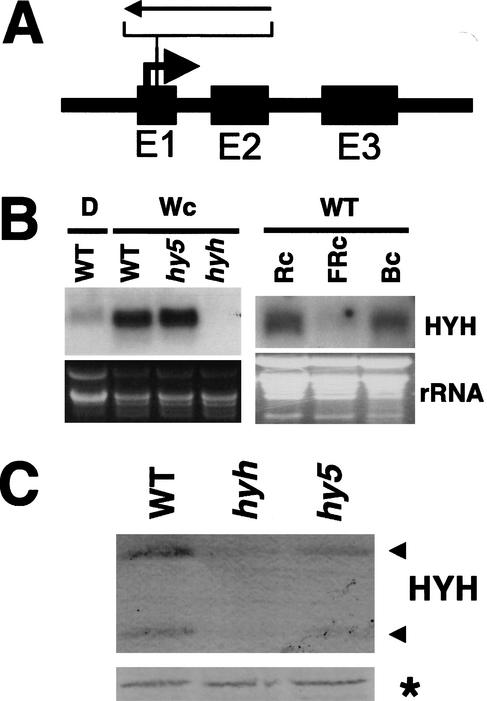
Figure 4.
Identification of a knockout mutation in the HYH gene. (A) Schematic illustration of the HYH gene organization with the position of the T-DNA insertion indicated. (B) Northern blot of 20 μg of total RNA prepared from 6-DAG dark-grown wild-type (WS) seedlings (D), and 6-DAG wild-type (WS), hy5-ks50, and hyh seedlings grown in white light, respectively. Right panel shows Northern blot of 20 μg of total RNA prepared from 6-DAG wild-type (Col-0) seedlings grown in continuous red (108 μmole/sec per m2), far-red (160 μmole/sec per m2), and blue (16 μmole/sec per m2) light, respectively. The HYH RNA is detected as a single band at ∼600 bp. (Bottom panels) The ethidium bromide-stained gel serving as a loading control. (C) Immunoblot of 10 μg of total protein prepared from 3-DAG wild-type (WS), hyh, and hy5-ks50 seedlings grown in white light. The two proteins detected with the HYH antibodies are indicated with arrows to the right. The asterisk marks a cross-reacting protein serving as a loading control.

