
Figure 1.
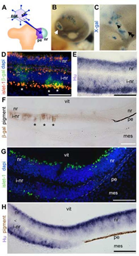
Pigmented epithelium transforms into neural retina and a non-neurogenic, non-pigmented tissue.
(A) Schematic of the operation for introducing FGF-expressing retrovirus into the pigmented epithelium. The retroviral vector co-expresses FGF and β-gal and is introduced into the pigmented epithelium domain of the optic vesicle. (B) Whole mount chick eye at e5 after infection as in A. Infected cells (blue: X-gal) found at the center of an area of depigmentation (arrow). (C) Magnified view of a depigmented patch, single arrow indicates center, double arrowheads indicate transition zone. (D) Immunohistochemistry on sections through center of infected portions of eye as in C (single arrow) with antibodies against islet-1 (red) and β-gal (green). (E) In situ hybridization on sections as in D were examined using cRNA directed against Hu (purple). (F) Immunohistochemistry on sections through the edges of infected portions of the eye as in C (double arrowhead), with antibody against β-gal (brown) to indicate foci of infection. At a distance from the infected cells, the induced neural retina transitions back into pigmented epithelium. (G) Transition zone as in F probed with an antibody against islet-1 (green) and DAPI nuclear stain. (H) Transition zone as in F expressing Hu (purple). Brown is endogenous pigmentation. pe: pigmented epithelium, nr: neural retina, vit: vitreal surface, i-nr: induced neural retina, mes: periocular mesenchyme. Scale: in D-G bar=50 μm; in H, I bar=100 μm.