
Figure 2.
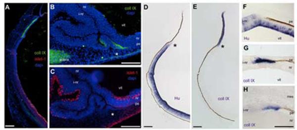
CollagenIX is expressed at the transition from pigmented to neural tissue.
Immunohistochemistry and in situ hybridization on e5 eye tissue. (A) Antibodies against collagenIX (green) and islet-1 (red) are expressed in unique domains. Blue is DAPI staining. (B) CollagenIX is ectopically expressed in the transition zone (asterisk). (C) Section adjacent to B, the transition zone in B is contiguous with an induced neural retina, identified by islet-1 (red). (D, E) HuD and CollagenIX expression on adjacent sections of normal eye, asterisks indicate mutually exclusive expression of HuD and collagenIX (F) Transition zone, HuD expression. (G) CollagenIX expression, adjacent section to F. (H) CollagenIX expression in a transition zone at the extreme posterior of the optic cup. L: lens, vit: vitreal surface, pe: pigmented epithelium, nr: neural retina, vit: vitreal surface, i-nr: induced neural retina, mes: periocular mesenchyme. Scale: in A-E bar=150μm; in F-H bar =50μm.