
Figure 5.
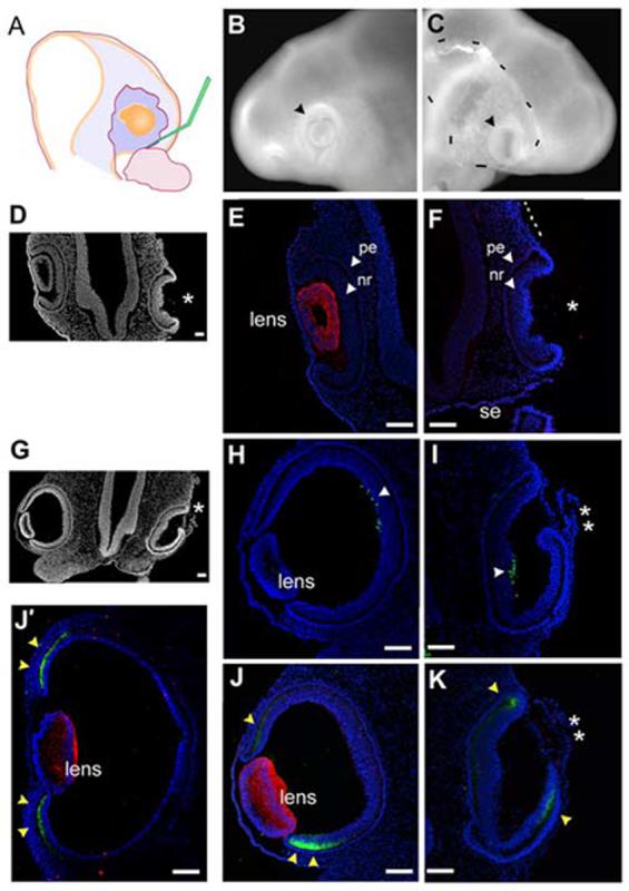
The optic cup can form without concomitant lens formation.
(A) Stage 12+ pre-lens ectoderm is surgically removed from over the optic vesicle. (B) Contralateral control eye, 24 hr after surgery (stage 16). Optic vesicle has formed a cup (arrowhead) with a centrally located lens. (C) Operated side, the vesicle has invaginated and no lens is apparent (arrowhead). Dashes indicate the edge of surface ectoderm, as it has not reformed over the optic tissue. (D) Section (coronal) through the eye region of embryo in B,C. Operated side formed an optic cup, no lens is seen (asterisk). (E) Delta-crystallin expression identifies lens tissue, optic cup morphology is seen with DAPI stained nuclei. (F) Delta-crystallin expression is not seen in the lens-less optic cup. The surface ectoderm is not present over the optic cup or the more dorsal portion of the head (dashed line). (G) Low magnification view of section through the eye region of an embryo removed 40 hr (stage 19) after pre-lens ectoderm removal at stage 12+. (H,I) Islet-1 expression (arrowheads) confirms that the lens-less optic cup has been specified correctly into pigment epithelium and neural retina. (J,K) Delta-crystallin (red) and collagenIX (green) expression on a section adjacent to G. Delta-crystallin is seen in the contralateral control eye (J), but the lens-less optic cup does not react (K); the tissue in front of the cup is not lens tissue (double asterisk). CollagenIX is more robust in the ventral than the dorsal domain at this stage (yellow arrowheads), but by stage 21 both dorsal and ventral domains are equal (J’). In the lens-less optic cup (K) collagenIX is also expressed, and in a dorsoventral pattern similar to that of the control eye. Scale: in A, D-E bar=100μm; in B,C bar=100μm.