
Figure 6.
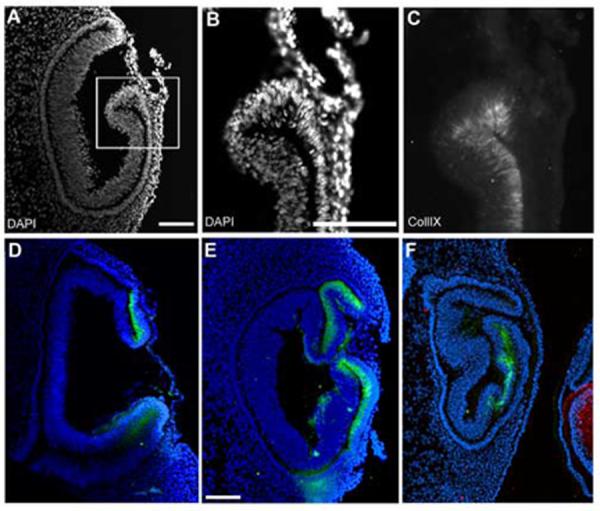
The ciliary body marker collagenIX is expressed in lens-less optic cups.
(A) DAPI stained nuclei reveal the morphology of a lens-less optic cup. (B,C) higher magnification of blocked area in A, with DAPI (B) and collagenIX (C) signal illustrates that collagenIX staining is specific for the inner layer of the optic cup, even in the absence of lens tissue. (D-F) Three additional examples of lens-less optic cups, all internally controlled with delta-crystallin staining in the contralateral eye, not shown, except for F, the control lens of an adjacent embryo. Scale: bar=100μm.